

Spine Treatment
Spine Treatment to Relieve Your Pain
Our Approach to Your Spine Health
CORE Orthopedics is dedicated to helping you get back to living a pain-free life. Whether that’s with minimally invasive surgical procedures or non-operative options, our team works with you to find the best choice to relieve your back pain.
Endoscopic Spinal Procedures at CORE Orthopedics
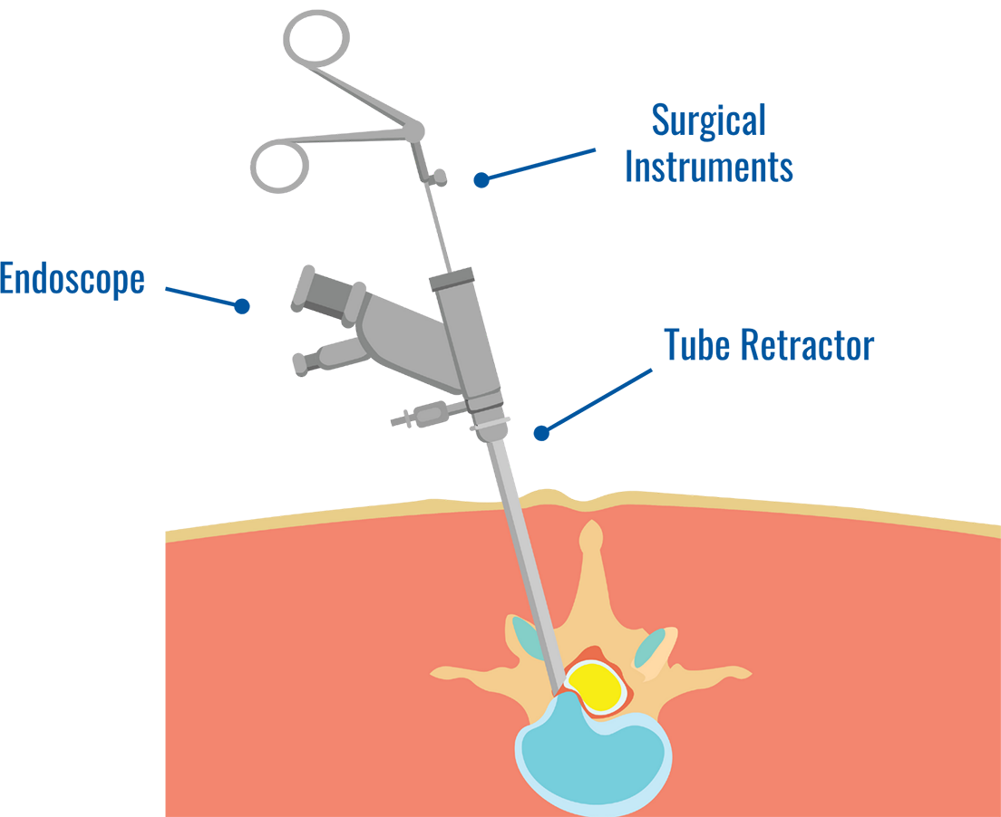
Dr. Joseph Krob specializes in endoscopic spine surgery which reduces recovery time and avoids long hospital stays.
He also provides more complex spine surgeries depending on a case by case basis.
This minimally invasive procedure uses a small camera inserted through a tiny incision to treat herniated discs, spinal stenosis, and nerve compression.
This procedure often results in less pain, smaller scars, and a faster recovery compared to traditional spine surgery.
Cervical (Neck) Spine Procedures
Cervical Spine Procedures focus on the neck to relieve nerve compression while stabilizing the upper spine. The goal is to eliminate pain that radiates into the head, shoulder and arms.
ACDF (Anterior Cervical Discectomy and Fusion)
Removes a herniated or degenerative vertebral disc in the neck and replaces it with a bone graft, relieving pressure to the spinal nerve roots.
Cervical Decompression
Removes elements pressing on the spinal cord or nerves in the neck, restoring sensation and function.
Cervical Arthroplasty (Disc Replacement)
Replaces a diseased disc with an artificial one to relieve pain and improve movement in the neck.
Thoracic (Mid-Back) Spine Procedures
Thoracic Spine Procedures stabilize the spine and decompress nerves in the mid-back region that cause pain.
Decompression
Relieves pressure on the nerves or spinal cord in the mid-back to stop pain and numbness.
Fusion
Permanently joins two or more vertebrae in the thoracic spine to provide stability and correct deformity.
Lumbar (Lower Back) Spine Procedures
Lumber Spine Procedures treat herniated discs, instability and more to resolve chronic lower back pain.
Minimally Invasive Decompression
Removes herniated discs and overgrown vertebral bone and soft tissue, to relieve nerve root compression in the lumbar spine.
Lumbar Fusion
Spinal fusion creates a solid mass of bone and stabilizes the spine.
Total Disc Arthroplasty
Replaces a damaged lumbar disc with an artificial disc to preserve mobility and reduce pain.
Anterior, Lateral, and Posterior Fusion
Access and stabilize the vertebrae from the front, side, or back, allowing for tailored care.
Non-Operative Spine Treatments
We always begin by exploring non-surgical methods to address your pain and restore function.
Lumbar Transforaminal Epidural Steroid Injections
An injection used to relieve lower back and radiating pain.
Kyphoplasty
Repairs a vertebral compression fracture, restoring the spines natural shape and relieving pain.
SI Joint Fusion
Stabilizes the sacroiliac joint to eliminate chronic lower back and buttock pain.
Dr. Joseph Krob
Dr. Joseph Krob is a Chicagoland native and completed his Orthopedic Surgery Residency at Loyola University Medical Center in Maywood, IL after earning his medical degree at the University of Illinois College of Medicine.
During his fellowship in spine surgery, Dr. Krob learned minimally-invasive spine decompression and fusion techniques, and has a passion for performing endoscopic spine procedures.
His clinical spine practice focuses on operative and nonoperative spine pathology such as stenosis, trauma/fractures, disc herniations, spondylolisthesis, and scoliosis among others.
Struggling with Back Pain? Call CORE Orthopedics Today.
Is back pain holding you back from living the life you deserve? Call CORE Orthopedics today and start living pain-free once again.
Testimonial

Back surgery
This video may contain descriptions or footage of medical procedures that are not suitable for all viewers. Viewer discretion is advised.
Learn More

Joseph Krob, MD
Dr. Krob is the newest member of the CORE Orthopedics team, joining in fall 2025 following completion of Orthopedic Spine Surgery Fellowship at Kaiser Permanente Northern California in Oakland, CA. He is Board Eligible for Orthopaedic Spine Surgery under the American Board of Orthopaedic Surgery (ABOS). Throughout his career, he has published numerous papers in a variety of peer-reviewed journals including North American Spine Society, American Journal of Sports Medicine. He maintains memberships to the North American Spine Society, American Academy of Orthopaedic Surgeons, and the Society for Minimally Invasive Spine Surgery. He is committed to life-long education by attending annual conferences to continuously provide evidenced-based, state-of-the-art treatment options for his patients. During his fellowship in spine surgery, Dr. Krob learned minimally-invasive spine decompression and fusion techniques, and has a passion for performing endoscopic spine procedures. His clinical spine practice focuses on operative and nonoperative spine pathology such as stenosis, trauma/fractures, disc herniations, spondylolisthesis, anThis video may contain descriptions or footage of medical procedures that are not suitable for all viewers. Viewer discretion is advised.
Location Information


Surgical Affiliations

Geneva Surgical Suites
119 Elizabeth Ln., Genoa City, WI 53128
Phone: 262-295-1213
Alexian Brothers Medical Center
800 Biesterfield Rd.
Elk Grove Village, IL 60007
Phone: 847-437-5500
St. Alexius Medical Center
1555 Barrington Rd.
Hoffman Estates, IL 60169
Phone: 847-843-2000
Advocate Good Shepherd Hospital
450 West Highway 22
Barrington, IL 60010
Phone: 847-381-0123